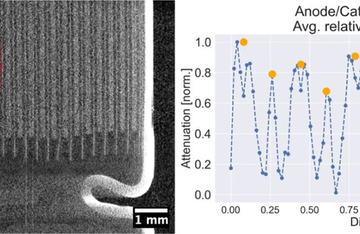
Although X-ray imaging covers a variety of experimental approaches, most of them carry the same basic requirements for a detector: high efficiency and good spatial resolution. A recently developed grating X-ray interferometer (Vila-Comamala et al. 2021) utilized these features of an EIGER2 detector for X-ray phase-contrast imaging in the biomedical field. The detector’s efficiency at energies from 15-25 keV allowed the exposure time to be shortened and harnessed most of the limited flux from a microfocus X-ray source.
The new EIGER2 family also includes high-energy sensors for even better efficiency in mid- and high-energy X-ray regimes, as well as two energy thresholds for suppressing cosmics.
Moreover, the EIGER2 detector series was a crucial component of the recently developed plenoptic multi-beam approach (Sowa & Korecki 2020; Sowa et al. 2020). In this setup, each beam projection results in a few photons per detector pixel, so in order to detect them, the detector also needs to suppress the high-energy photons through a high-energy threshold. In the subsequent averaging procedure, the individual projections need to be aligned precisely, and this requires a detector with a sharp point-spread function and small pixels – like EIGER2.
- Achieve high efficiency for a wide range of X-ray energies with a choice between two sensor materials – silicon (Si) and cadmium telluride (CdTe).
- Our detectors feature a high dynamic range, with noise-free and offset-free readout.
- Signals are highly resolved due to a one-pixel-wide point-spread function.
X-ray Imaging Radiography and CT in Laboratories
- Make digital spectrum adjustments thanks to EIGER2 detectors’ dual-energy discrimination.
- Work with extremely low-flux sources thanks to EIGER2 detectors’ readout, which is free of dark signals and noise.
- Take faster measurements with EIGER2’s continuous readout.


